

MAGNETIC RESONANCE IMAGING (MRI)
WHAT IS MAGNETIC RESONANCE IMAGING (MRI)?
Magnetic Resonance Imaging (MRI) is a diagnostic imaging tool that uses a magnetic field and radio waves to generate detailed images of organs and tissues inside the body.
REASON FOR THE PROCEDURE
When physicians are not able to make a complete diagnosis from a physical exam, MRI is used to provide a noninvasive view of internal organs, tissues and bone. MRI is the most frequently used procedure for viewing the brain and spinal cord, the chambers of the heart and blood vessels, masses in organs and tissues and joint, disk and bone abnormalities.
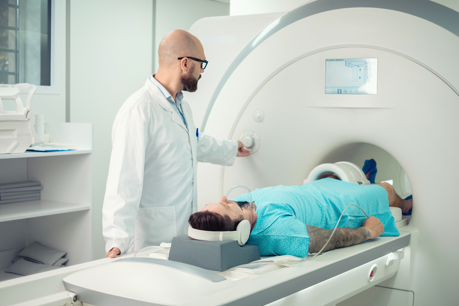
THE PROCEDURE
The procedure requires you to lie inside a large cylinder while multiple images are taken and transferred electronically to a computer. The images created are very detailed and show tissue and organ alignment in the body. Any swelling or enlargements, masses or other abnormalities can be detected.
POTENTIAL SIDE EFFECTS, RISKS, AND COMPLICATIONS
Because MRI uses powerful magnets, having any metal in your body can be an issue. Even if not attracted to the magnet, metal can distort the MRI image. Metal items that may interfere with MRI include metal heart valves, heart defibrillators, internal infusion pumps, pacemakers, hearing implants, intrauterine devices, screws or plates used to stabilize bone or metal from a bullet or shrapnel. At Excel Imaging, your Esse Health Radiologist and staff will discuss the procedure with you in more detail and answer any questions you may have.